
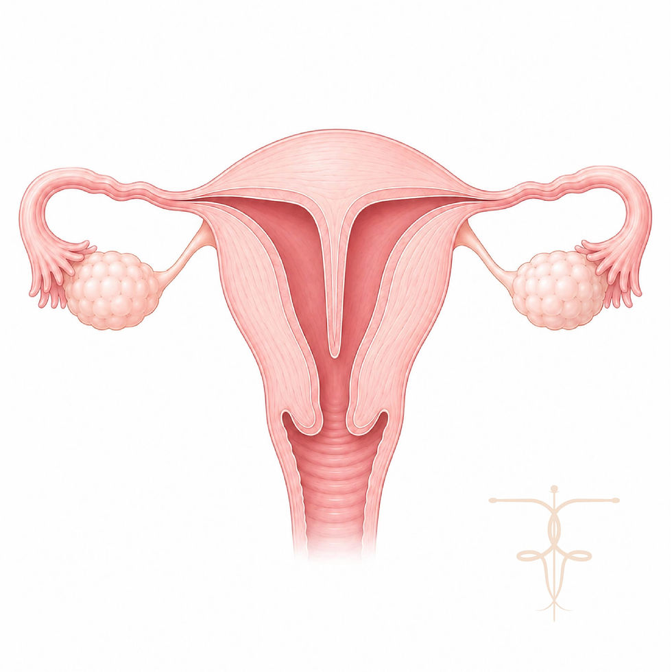
Uterine Septum
A uterine septum (septate uterus) is a congenital condition where a band of tissue divides the inside of the uterus partially or completely. It develops before birth when the uterus does not form normally during fetal development. Many women are unaware they have a uterine septum until they experience fertility difficulties, recurrent miscarriage or pregnancy complications.
Commonality
Uterine septa are estimated to affect around 5–10% of women and are among the most common congenital uterine abnormalities.
Causes
A uterine septum develops before birth when the two structures that normally fuse to form the uterus do not completely merge.
As a result:
A thin wall of tissue remains inside the uterus
The uterine cavity becomes partially or completely divided
Types of uterine septum include:
Partial septum, which extends from the top of the uterus but does not reach the cervix
Complete septum, which extends from the top of the uterus to the cervix and may occasionally extend into the vagina
Symptoms
Many women have no symptoms and the condition is often discovered during fertility investigations.
Possible symptoms include:
Difficulty conceiving
Recurrent miscarriage
Preterm labour
Abnormal fetal position during pregnancy
Painful periods
Occasional abnormal bleeding
Effects on Fertility
A uterine septum does not always prevent pregnancy, but it can increase the risk of fertility and pregnancy complications.
Possible effects include:
Reduced implantation success
Recurrent miscarriage
Preterm birth
Malpresentation of the baby
Increased pregnancy complications
Approximately many women are still able to conceive, and surgical treatment may improve pregnancy outcomes, particularly after recurrent miscarriage.
Diagnosis
Diagnosis requires detailed imaging to assess the shape of the uterus.
Investigations may include:
3D transvaginal ultrasound
Saline infusion sonography (SIS)
MRI scanning in complex cases
Diagnostic hysteroscopy
A 3D ultrasound scan is particularly useful because it provides detailed views of the uterine cavity and helps distinguish a septate uterus from other uterine abnormalities.
Treatments
The standard treatment for a uterine septum is hysteroscopic septum resection where a small camera is inserted into the uterus and the septum is carefully removed under direct visual guidance.
This minimally invasive procedure:
Removes the dividing tissue inside the uterus
Requires no external incisions
Is usually carried out as a day-case procedure
Preserves fertility
Allows rapid recovery
The procedure aims to restore a normal uterine cavity and improve reproductive outcomes.
Ongoing Care
Ongoing care after treatment may include:
Follow-up scans to confirm healing
Short-term hormonal treatment where recommended
Review of symptoms and fertility plans
Guidance on when to begin trying for pregnancy
Women are often advised to wait around two months after surgery before attempting pregnancy.
Living with the Condition
Many women with a uterine septum have no symptoms and may only discover the condition during fertility investigations. However, recurrent miscarriage and fertility difficulties can be emotionally distressing.
Women may experience:
Anxiety about future pregnancies
Grief following pregnancy loss
Uncertainty during fertility treatment
Compassionate care, clear explanations and appropriate treatment can provide reassurance and improve confidence when planning future pregnancies.
When to See a Specialist
You should consider specialist assessment if you:
Have experienced recurrent miscarriage
Are having difficulty conceiving
Have experienced pregnancy complications without a clear explanation
Have been told you may have a uterine abnormality
Have symptoms suggestive of a congenital uterine condition
Early diagnosis and treatment can improve reproductive outcomes and support future pregnancy planning.